Saturday, May 1, 2010

Immunological variants in children

Children have an immune system that differs from that of adults in several ways; in young children it is fairly immature and cannot provide adequate protection since development requires repeated exposure to new antigens. Several factors can affect the development of a child’s immune system as illustrated in figure 2. A good example for this statement is the results of a recent study conducted at Duke University Medical Centre in relation to peanut hypersensitivity in children. A controlled dose of peanuts was given daily to the patients as the treatment. “At the start of the study, these participants couldn't tolerate one-sixth of a peanut, six months into it, they were ingesting 13 to 15 peanuts before they had a reaction”. 8 These results are possible due to immunological changes, like desensitization, that make adaptation and tolerance possible in both children and adults.8 An example of this can be seen in the use omalizumab (xolair) to treat moderate to severe persistent allergic asthma caused by allergens in the air. Xolair helps reduce the amount of asthma attacks. 9

Neonates are considered to be in a state of immunodeficiency. During the first months of their lives they obtain immunologic protection from their mothers in the form of immunoglobulins (IgG and IgA) that had crossed the placenta in utero, and through breast milk post utero4. Breast milk contains macrophages, lymphocytes, granulocytes, antibodies, complement and enzymes which destroy bacteria. Yet, these immunoglobulins are insufficient protection against viral and intra-cellular infections such as cytomegalovirus, Herpes simplex, Listeria and Toxoplasma. It has been shown that the maternal immune system can directly affect the child’s hypersensitivity. An example of this is that maternal cells have been found in an abnormally high number of patients with Juvenile dermatomyositis and these cells contain the mothers HLA that is thought to start the autoimmune response. 7 Children who suffer from this muscle damaging disease may also suffer organ failure.

Studies have also shown that newborns posses a depressed number of cells involved in the adaptive immune response and the C3 component of the complement system.3 In a study done in 2006, researchers found low numbers of CD3+ T cells and a decreased level of cytokine activity, which are essential for initiation of the adaptive immune response in neonates.1 Overall, neonates are more susceptible to viral and intracellular infections due to a deficiency in NK cells and cytotoxic T cells. Although neonates posses functional lymphocytes by around two months of age their antibody production does not reach adult levels until the age of four. This deficiency results in high mortality rates as a result of infection. New research aims to find methods to reduce the morbidity and mortality rates of newborns by reinforcing their dwindling immune system.4

Once a child hits puberty, the increasing amounts of gonadotropic hormones induce the secretion of androgens and estrogens, which affect the immune response. Estrogens increase IgG and IgM secretion by mononuclear cells while testosterone may suppress the stress response associated with infection. Another important change that occurs after puberty is the “involution of the thymus”. 6 Thymic involution causes a decrease in naïve T lymphocytes resulting in the formation of T cells that are responsive to foreign antigens and have yet to be stimulated by foreign substances. Also, it is believed that involution of the thymus helps to protect the body from autoimmunity disorders.


Questions:

1. During the first months of their lives neonates obtain immunologic protection from:

a. Live vaccines

b. Intravenous infusions of immunoglobulins

c. Blood transfusions

d. Mothers immuglobulins pre partum and breast milk post partum

2. Which immunoglobins are secreted due to estrogen secretion?

a. IgM and IgE

b. IgG and IgM

c. IgG and IgA

d. IgA and IgE

3. Neonates posses functional lymphocytes at about 2 months of age, their antibody production does not reach adult levels until the age of

a. 2

b. 4

c. 10

d.15

4. What environmental factors can affect the development of the immune system?

5. When a child reaches puberty, the secretion of Estrogen causes mononuclear cells to increase production of which two immunoglobulins?

6. Deficiency in which two types of cells would attribute to neonates increased susceptibility to viral and intracellular infections?

References:

1. Engelmann, Ilka *; Moeller, Ulrike; Santamaria, Andrea; Kremsner, Peter G.; Luty, Adrian J.F. Differing activation status and immune effector molecule expression profiles of neonatal and maternal lymphocytes in an African population. Immunology. December 2006; 119(4):515-521

2. Lott, Judy Wright. State of the Science: Neonatal Bacterial Infection in the Early 21st Century. Journal of Perinatal and Neonatal Nursing. January/March 2006; 20 (1): 62-70

3. Firth, Matthew A.; Shewen, Patricia E.; Hodgins, Douglas C. Passive and active components of neonatal innate immune defenses. Animal Health Research Reviews. December 2005; 6 (2): 143-158

4. Ohlsson A, Lacy J. Intravenous immunoglobulin for suspected or subsequently proven infection in neonates. Cochrane Database Syst. Rev. 2004

5. Jaspan, H.B., Lawn, S.D., Safrit, J.T., et. al.; The Maturing Immune System: Implications for Development and Testing HIV-1 Vaccines for Children and Adolescents. AIDS. 2006; 20:483-494. http://www.medscape.com/viewarticle/524225 . April 10, 2010.

6. Eric S. Lugada, Jonathan Mermin, Frank Kaharuza, Elling Ulvestad, Willy Were, Nina Langeland, Birgitta Asjo, Sam Malamba, and Robert Downing. Population-Based Hematologic and Immunologic Reference Values for a Healthy Ugandan Population. Clin. Diagn. Lab. Immunol., Jan 2004; 11: 29 - 34.

7. Reed, A.M., McNallan, K., Wettstein, P., Vehe, R., and Ober, C. (2004). Does HLA-dependent chimerism underlie the pathogenesis of juvenile dermatomyositis? Journal Immunology; 172, 5041-5046.

8. Burks W. ;M.D. Studies show children can complete treatment for peanut allergies and achieve long-term tolerance. From www.dukemednews.org 2009. Available at: http://www.sciencecentric.com/news/article.php?q=09032026-studies-show-children-can-complete-treatment-peanut-allergies-achieve-long-term-tolerance. Accessed April 14, 2010.

9. 2010 Genentech USA, Inc. and Novartis Pharmaceuticals Corporation. www.xolair.com. Retrieved April 29th 2010.

Figure 2: Storey M, Jordan S (2008) An overview of the immune system. Nursing Standard. 23, 15-17, 45-56.

IMMUNOSTIMULANTS

Immunostimulants are substances (drugs or nutrients) that increase the ability of the immune system to fight infection and disease (USIH). Typically, immunostimulants are divided into specific and non-specific categories. Specific imunostimulants stimulate an immune response to one or more specific antigenic types. This highly specific adaptive response begins when the patient is infected for a first time. The immune system reacts to the new pathogen and develops permanent protection by T and B lymphocytes. These immune cells will protect the body when subsequently met by the same pathogen. This immune response is mounted when macrophages engulf the foreign antigens and present them in the lymph nodes. These antigens are then recognized by T-helper, T-Killer, or B cells. B cells enlarge to produce plasma cells that produce antibodies, impeding the microbes and marking them to be engulfed by macrophages. When the virus is removed, memory cells are formed by T- and B cells so when there is re-exposure a quick response ensues. Vaccines, another type of specific immunostimulant, also mimic natural infection preparing the system for a quick response to re-exposure (Figure 1) Adjuvants also enhance the response of the immune system specifically. Some mechanisms of action for adjuvants through noncovalent binding are: prolonging the stimulus through a delay in the release of immunogen; enhancing the uptake of immunogen by antigen-presenting cells; inducing co-stimulatory molecules; and stimulating the Toll-like receptors on the surface of macrophages, which in turn induces the cytokine production that enhances the response of T cells and B cells to the antigen (Levinson).

Non-specific immunostimulants do not have any antigenic specificity, but rather act as general stimulants that enhance the function of certain types of immune cells. In non-specific immunostimulation, a primary augmentation of the immune response is by the anatomical and mechanical barriers that are associated with chemical and biological agents. (Medzhitov, R., Janeway, C. 2000). Cytokines, or interleukins, mediate non-specific immunostimulation signal by affecting cells’ behaviors. In addition to inducing growth, differentiation, and apoptosis, the upregulation of these mediators increase the immune response and helps to contain different infectious agents (Vinderola, C. et al. 2006). Cytokines therefore provide a strong defense in several clinical cases that would potentially cause serious damage to the host. For example, granulocyte-macrophage colony stimulating factor (GM-CSF) is a cytokine secreted by macrophages, T cells, mast cells, endothelial cells and fibroblast that effectively stimulates stem cells to produce granulocytes and monocytes (Crawford J. 1991). GM-CSF’s are used as medications to stimulate the production of white blood cells following chemotherapy. This non-specific immunostimulant has also been considered a potential vaccine adjuvant in HIV-infected patients and is waiting for FDA approval (Lieschke GJ. 1992).

Endocrine hormones such as PRL, GH and thyroid hormones have been demonstrated to have a role in immunostimulation as well. These hormones exert their effect indirectly as anabolic and stress-modulating hormones affecting cells of the immune system (Dorshkind and Horseman 2000). These endocrine signals modulate immunosuppression associated with glucocorticoids. The positive effects of these hormones on the immune system occur as an adaptation to stress (Figure 2); therefore, they are not required for the generation of an immune response in healthy individuals (Dorshkind and Horseman 2000). Immunostimulants are also found in the form of food derivates. Vitamins, minerals, and fatty acids generate a proliferation in antigen-specific antibody (Figure 3). Vitamins, minerals, oligosaccharides and lactic bacteria also augment T cells and amplify their propagation during an immune reaction and elevate phagocytic and NK cells action (Shuichi, K. and Masanobu, N. 2004).

FIGURES:

Fig 1: Mechanism of action of vaccines (www.cdc.gov)

Figure 2: The interaction between stress and the endocrine system are highlighted here. Glucocorticoids (CORT) have immunosuppressive effects via glucocorticoid receptor (GR) mechanisms as seen on the left. This results in maladaptive response in the immune system. On the right, the expression of prolactin (PRL) and/or growth hormone (GH) on immune cells interact with GR preventing the interaction of STAT and GR, minimizing the negative effects of glucocorticoids on immune cells. (Dorshkind and Horseman 2000)

Figure 3: Manipulation of the immune system by food products. The two systems affected are the innate and adaptive immune system. Lactic acid and vitamins have a direct effect on the innate system by increasing phagocytic activity and NK cells. Vitamins, minerals, fatty acids and oligosaccharides affect the adaptive immune system by increasing T cell response and antibody production. (Shuichi, K. and Masanobu, N. 2004)

CLINICAL CASES:

1. GI TRACT INFECTION: Normal Intestinal Microbiota competition against invading pathogen (Cytokine Interaction in Immunostimulation)

CD is a very healthy woman. Her favorite breakfast is a parfait, with yogurt, fresh fruit and granola. She has it every morning after her daily run. For her birthday she decides to go out for dinner with a couple of friends. She had some pork with mashed potatoes. After a few hours she starts getting abdominal pains and she feels bloated. She has both urinary and fecal incontinence, flatulence and experiences vomiting. She can’t even drink water. She visits her physician and he diagnoses her with food poisoning. He orders her to have small amounts of yogurt and Gatorade to prevent dehydration. After 2 days she is feeling much better. She can eat solid food again and her bowel movements have normalized.

Figure 4: Illustration of the natural defense systems of the intestine (www.customprobiotics.com). The intestine, composed of villi and crypts coated with mucus. At the bottom of the crypts lie Paneth cells that release antimicrobial molecules into the gut lumen. The intestinal flora, present mainly in the colon, forms a natural barrier to pathogens. The intestinal immune system comprises cells disseminated beneath the epithelium, lymphocytes, both B and T. When a lymphocyte is activated, it leaves the mucosa in lymph and enters the bloodstream via the thoracic canal eventually colonizing the same mucosa or other mucosal effector sites.

EXPLANATION: Fermented dairy products (ex. yogurt) contain specific metabolites such as peptides and exopolisacharides that enhance B cell proliferation and increase the secretion of Ig A and Ig G antibodies in the gut mucosa. This leads to prevention of infection from bacteria and other microbes in the GI tract. In recent studies, PMFKM (kefir microflora) proved to increase a number of cytokines such as IL-4, IL-6, IL-10, IL-12, IFNc and TNFa mostly in the small intestine. Cytokine secretion was also observed in blood serum, reflecting the inmunostimulant properties of the PMFKM. This demonstrates a manipulation of the constituents of the intestinal lumen through dietary means, enhancing the health status of the host.

2. Non-specific Immunostimulation Treatment

A 30 year-old man that is a cook was sharpening his cooking knives when one slipped and made a 2.5 inch incision in his arm. He rushes to the hospital because he can’t stop the bleeding. The doctors gave him stitches. After a week, he returns to the hospital because the injury is infected. His CBC shows that he has a low WBC count for someone with an infection. The physician asked if he had omitted anything from his medical history that could explain the abnormal results of the infection. He admitted to being HIV positive. Knowing now that this patient is immuno-compromised, the physician opened the wound and flushed it with WFI and gave him an infusion of tetrachlorodecaoxygen in order to stimulate a proper macrophage phagocytic response. After 2 weeks, his wound healed and he was able to go back to work.

Fig 5: Mechanism of action of WF 10 with chronic inflammation. (biomaxx-sys.com)

Explanation: Tetrachlorodecaoxygen has been commonly used for the treatment of external wounds. WF 10 is an intravenous infusion based on the tetrachlorodecaoxygen (TCDO) drug that stimulates phagocytosis by macrophages (Penpattanagul, 2007). WF 10 inhibits proliferation, IL-2 production of anti-CD3 stimulated PBMC, and the nuclear translocation of NFATc (transcription factor); WF 10 induce cytokines like IL-1β, IL-8, and TNF-α (Giese, 2004). WF 10 was developed as an adjunctive therapy to simultaneously combat antiretroviral and opportunistic infection prophylaxis in AIDS patients. It was also approved by the Thailand government for use in post radiation cystitis in cervical cancer patients (Drugs RD, 2004).

3. Prophylactic treatment against febrile neutropenia in cancer patients

A patient who was recently diagnosed with cancer is undergoing chemotherapy. As a consequence of this therapy, he develops bone marrow suppression. Within a few days, he develops a febrile neutropenia. A CBC was ordered showing a decrease in neutrophils. The doctor, not wanting to remove him from chemotherapy treatment, prescribes him filgrastim, a G-CSF treatment to reduce his febrile neutropenia. After a week of treatment, the patient goes for a follow up visit. He tells the doctor that he has not had anymore fevers. A follow-up CBC showed that his WBC levels had increased.

Fig. 6: The role of the neutrophil consist of their response to foreign invaders. Once the invaders enter the blood vessels, neutrophils detect them and commence phagocytosis in order to remove the bacteria from the system. (hivandhepatitis.com)

Fig. 7: Administration of G-CSF into the body travel to the bone marrow where it stimulates the formation of granulocytes leading to an increase of neutrophils in the blood system. (probiomed.com.mx)

EXPLANATION:

Cancer patients undergo chemotherapy therapy in order to decrease the tumor burden and prolong survival of the patient. Tumors are more susceptible than normal tissue to chemotherapeutic agents because they have a higher proportion of dividing cells. However, because certain types of normal tissue have a high rate of division rate like the bone marrow, mucosa and epidermis, they are also susceptible to the effects of chemotherapy (Meric-Bernstam). Therefore, treatment with chemotherapeutic agents can produce noxious effects, such as bone marrow suppression, which leads to febrile neutropenia. Other side effects of chemotherapy include stomatitis, ulceration of the GI tract, and alopecia (Meric-Bernstam).

Febrile neutropenia increases infection-related morbidity and mortality; therefore, it is a significant dose-limiting toxicity in cancer treatment. (Aapro, 2006). Cancer patients developing severe or febrile neutropenia (FN) during chemotherapy frequently receive dose reductions and/or delays to their chemotherapy. A reduction or delay in therapy may impact the success of treatment, particularly when treatment purpose is either curative or to prolong survival. Recent analyses have shown that by reducing the risk of FN and chemotherapy dose delays and reductions there is a potential enhancement in survival rates for cancer patients. In order to reduce febrile neutropenia in patients undergoing chemotherapy, there has been an increase in the prophylactic use of granulocyte colony stimulating factors (G-CSFs), such as filgrastim, lenograstim or pegfilgrastim. G-CSFs influence the development of the neutrophils, acting as immunostimulants. G-CSF therapy will increase the level of neutrophils and shorten the length of the cycle sufficiently to prevent recurrent bacterial infections (Hammond et al., 1989).

___________________________________________________________________________

QUESTIONS:

1. What is the mechanism by which Prolactin (PRL) modulates the immune system during adaptive stress?

Answer: Prolactin on immune cells interacts with GR preventing the interaction of STAT and GR, minimizing the negative effects of glucocorticoids on immune cells. (Fig. 3)

2. True or False: Lactic acid bacteria and vitamins affects the adaptive immune system by increasing phagocytic activity and NK cells while Vitamins, minerals, fatty acids, and oligosaccharides affect the innate immune system by increasing T cell response and antibody production.

Answer: FALSE. Lactic acid bacteria and vitamins affects the innate immune system by increasing phagocytic activity and NK cells while Vitamins, minerals, fatty acids, and oligosaccharides affect the adaptive immune system by increasing T cell response and antibody production.

­­­3. What are the benefits of the prophylactic treatment against febrile neutropenia in cancer patients?

Answer: It stimulates the formation of granulocytes and eventually increasing the levels of neutrophils in the body.

4. Adjuvants differ from protein carriers in that:

a. form covalent bonds with the immunogen

b. accelerate the release of the immunogen

c. diminish the immune response to an antigen

d. do not form covalent bonds with the immunogen

5. Which line of blood cells is the principal target when G-CSF or GM-CSF is administered?

a. Neutrophils

b. Lymphocytes

c. Red Blood Cells

d. Platelets

6. Once the adaptive immune system is activated, it can trigger the T-cell-mediated immune response and the antibody response, both are carried out by ______________.

A) T lymphocytes

B) B lymphocytes

C) Dendritic cells

D) Effector T cells

E) A and B

REFERENCES:

Crawford J, Ozer H, Stoller R, Johnson D, Lyman G, Tabbara I, et al. Reduction by

granulocyte colony-stimulating factor of fever and neutropenia induced by

chemotherapy in patients with small-cell lung cancer. New England Journal

of Medicine. 1991;325:164-70.

Dorshkind, K., & Horseman, N.D. The roles of prolactin, growth hormone,

insulin-like growth factor-1, and thyroid hormones in lymphocyte

development and function: insights from genetic models of hormone and

hormone receptor deficiency. Endocrine Reviews, 2000, 21(3), 292-312.

Hammond, W.P. IV, Price, T.H., Souza, L.M., and Dale, D.C. Treatment of cyclic

neutropenia with granulocyte colony-stimulating factor. N. Engl. J.

Med., 1989, 320:1306–1311.

Kaushansky Kenneth, Kipps Thomas J, "Chapter 53. Hematopoietic Agents: Growth Factors,

Minerals, and Vitamins" (Chapter). Brunton LL, Lazo JS, Parker KL: Goodman &

Gilman's The Pharmacological Basis of Therapeutics, 11e:

http://www.accessmedicine.com.lib.sanjuanbautista.edu:194/content.aspx?aID=95212

Levinson W, "Chapter 57. Immunity" (57). Levinson W: Review of Medical

Microbiology and Immunology, 10e: http://www.accessmedicine.com/content.aspx?aID=3334292

Lieschke GJ, Burgess AW. Granulocyte colony-stimulating factor and granulocyte-

macrophage colony-stimulating factor (1 and 2). New England Journal of

Medicine. 1992;327:28-35,99-106.

Lokeshwar, S.A. Febrile neutropenia in haematological malignancies

J Postgrad Med. 2005;51 Suppl 1:S42-8.

Medzhitov, R, Janeway, C. Innate Immunity. Advances in Immunology. August 3,

2000. Vol.343:338-44. Num. 5.

Meric-Bernstam Funda, Pollock Raphael E. Brunicardi FC, Andersen DK, Billiar TR, Dunn

DL, Hunter JG, Matthews JB, Pollock RE: Schwartz's Principles of Surgery, 9e:

http://www.accessmedicine.com.lib.sanjuanbautista.edu:194/content.aspx?aID=50211

63.

National Institute of Allergy and Infectious Diseases. (August 25, 2008). How Vaccines

Work. Retrieved from

http://www.niaid.nih.gov/topics/vaccines/understanding/Pages/howWork.asp

Shuichi, K. and Masanobu, N. Modulation of Immune Functions by Foods. October,

2004. eCAM. 1(3) 241-250.

United States Institute of Health: National Cancer Institute.

Vinderola, C.G.; Perdigon, G.; Duarte, J.; Farnworth, E.; Matar, C. Effects of the oral

administration of the products derived from milk fermentation by kefir

microflora on the immune stimulation. Journal of Dairy Research. Cambridge

University Press, England. 2006. Vol.73 n.4 vol.73. p.472-479.

Interaction Between the Central Nervous System and the Immune System

The interaction between the Central Nervous System (CNS) and the Immune System is critical to maintaining normal health. A fundamental aspect of this interaction is the presence of the Blood Brain Barrier (BBB), the physical separation between the vascular system and the tissues of the brain and spinal cord. The BBB works to prevent the diffusion of toxins, bacteria, antibodies and large proteins from crossing the barrier while simultaneously allowing the passage of small hydrophobic substances such as O2, CO2 and hormones in cerebrospinal fluid (CSF)[1].

Microglia act as the first line of active immune defense for the CNS. In the brain and spinal cord, microglia work as phagocytes to remove damaged neurons, plaques and infectious agents. Antigenic activation of microglia may also stimulate an increase in astrocyte activity and vice-versa. Together, astrocytes and microglia differentially regulate trafficking of lymphocyte subsets across brain endothelial cells[2].

A second mode of interaction between the CNS and the immune system occurs when an antigen crosses the BBB and infiltrates nervous tissue. In order to defend against such an attack, the CNS has countermeasures in place to halt and eliminate antigens. Effector and memory T cells constantly survey the CNS to protect from infection and the manifestation of cancer[1]. This neuroinflammatory response, if unregulated, may be detrimental to the CNS[1,3].

Upon injury to the CNS, neuroinflammation leads to infiltration of CD4 activated cells survival signals for T cells. During the inflammatory response, TGF-β secreted by astrocytes and glial cells is joined by IL-6 or IL-21 to form T-cells (TH-17). These T cells produce cytokines such as IL-17, IL-17F and IL-22 to regulate the inflammatory response within the CNS[1,3].

A third mode of interaction between the CNS and immune system is the effect of hormones and cytokines on overall immune function. The immune response triggers the Hypothalamic-Pituitary-Adrenal axis (HPA axis), which initiates the release of glucocorticoids such as cortisol. Cortisol limits the cytokine induced inflammatory response and prevents an overreaction from the immune system. This chain of events has been interpreted to indicate that the immune system can act as a sensory system, notifying the brain of the presence of a threat and triggering a classical stress response[4].

Of the large number of known cytokines, the most relevant to the nervous system are IL-1, IL-6, TNFa, and the interferons. IL-1 is a considerably more potent activator of adrenocorticotropic hormone (ACTH) and glucocorticoid secretion than CRF itself[4]. Cytokines within the CNS activate immune responses that elicit tissue repair or cell damage and cytotoxicity resulting in necrosis and loss of oligodendrocytes[1,4].

The interactions between the CNS and immune system are very complex. Signaling by the HPA axis and its effects over the CNS and immune system are involved in regulating interactions between immune cells, their substances, and the CNS. A delicate balance must be maintained between the aforementioned components of the immune system and CNS in order to ensure a quality state of health for the patient.

Clinical Applications

1. Multiple Sclerosis

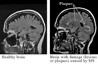
Multiple sclerosis is a chronic inflammatory disease characterized by recurrent episodes of demyelination that disturbs bundles of nerves in the white matter of the central nervous system. Components of the CNS affected by the presence of sclerotic plaques include the cerebellum, spinal cord, and projections of optic nerve, somatosensory pathways, costicospinal tract and basal ganglia [see fig. 2]. The cause of this disease is unknown but it is believed that it could be a T cell-mediated autoimmune disease caused by a combination of factors including heredity, susceptibility to infectious agents, production of autoantibodies by autoreactive B-cells against myelin antigens such as myelin basic protein, myelin oligodendrocyte glycoprotein and proteolipid protein [5]. In addition, increased frequency of developing the disease is associated with individuals that carry the HLA-2 or HLA-3 genes, genes that encode receptors for IL-2 or IL-7 [6], as well as relative lack of vitamin D and exposure to cold and temperate climates [6]. The symptoms are characterized by partial or complete loss of vision, muscle weakness, fatigue, numbness and problems with balance, speech, and generalized motor coordination, including bowel and urinary control [7, 9] [see fig. 1]. In 50% of patients the cumulative sensory and motor losses may lead to generalize muscular paralysis, thus requiring help for walking within 15 years of initial occurrence of symptoms [9]. Even though there is no cure, treatments involving administration of corticosteroids and interferon β-1a or β-1b have shown to reduce the progression of the disease.


Figure 1

(Multiple Sclerosis Symptoms. Health Kut. http://healthkut.com/blog/wp-content/uploads/2010/02/multiple_sclerosis-Symptoms.png. Accessed April 29, 2010.)

Figure 2

(Brain Scan Lesion Damage. Health.com. http://www.health.com/health/static/hw/media/medical/hw/h9991221.jpg. Accessed April 29, 2010.)

2. Myasthenia Gravis

Myasthenia gravis is an autoimmune disease where antibodies produced by T and B-lymphocytes reduces the number of functional nicotinic acetylcholine (Ach) receptors in the postsynaptic membrane of the neuromuscular junction [see fig.4]. This causes a decrease of response of the muscle fibers to Ach, thus eliciting a progressive muscular weakness. It has been established that molecular mimicry plays an important factor in this disease due to structural similarities in amino acid sequences shared with certain poliovirus proteins. Also, individuals carrying HLA-DR3 gene have increase risk of expressing the disease [11]. Clinical features of Myasthenia Gravis involve general muscle weakness that greatly manifests in the muscles of the arms, head and chest [9]. It characterizes by fatigue involving muscles of the eyes and eyelids, causing ptosis [see fig. 3], facial expression and of the pharynx, leading to problems with mastication, swallowing, holding the head upright and in speech [8, 9]. In severe cases of the disease, muscular weakness of the diaphragm and abdominal intercostal muscles produces respiratory paralysis, with a mortality rate of 5-10% [9]. It has been reported that 70% of patients with this disease have an abnormal thymus, therefore thymectomy, as well as administration of corticosteroids and immunosuppressant therapy is recommended [8, 9, 11]. However, an important therapeutic approach involves the administration of anticholinesterase drugs, such as neostigmine, which reduces the activity of cholinesterase, thus allowing the rising of Ach at the synapse in order to stimulate the existing receptors and producing muscle contraction.

Figure 3

(Ptosis (Drooping Eye). ADAM. http://wendyusuallywanders.files.wordpress.com/2008/01/mgdroop.jpg. Accessed April 29, 2010.)

Figure 4

(Myasthenia Gravis. Journal of the American Medical Association. http://jama.ama-assn.org/content/vol298/issue1/images/medium/jmn70071fa.jpg. Accessed April 29, 2010)

3. Lupus

Systemic Lupus Erythematosus (SLE) is an autoimmune disorder that is characterized by a multisystem microvascular inflammation with the generation of autoantibodies. The precise reason for the abnormal autoimmunity that causes lupus is not known. Patients with lupus produce abnormal antibodies in their blood that target tissues within their own body rather than foreign infectious agents. Many immune disturbances, both innate and acquired, occur in SLE, as illustrated in below[10].

Figure 5

(Bartels C, MD. Systemic Lupus Erythematosus. 2009. Medscape http://emedicine.medscape.com/article/332244-overview.)

It has been suggested that the development of autoantibodies involves a defect in apoptotic cells, it has been demonstrated that there is clustering of lupus autoantigens in the surface blebs of apoptotic cells. Thus, intolerant lymphocytes begin targeting normally protected intracellular antigens [10].

Lupus can cause disease of the skin, heart, lungs, kidneys, joints, and/or nervous system. Immune complexes form in the microvasculature, leading to complement activation and inflammation. Moreover, antibody-antigen complexes deposit on the basement membranes of skin and kidneys. In active SLE, this process has been confirmed based on the presence of complexes of nuclear antigens such as DNA, immunoglobulins, and complement proteins at these sites. Serum antinuclear antibodies are found in virtually all individuals with active SLE, and antibodies to native double-stranded DNA are relatively specific for the diagnosis of SLE[10].

Questions

1. What is the role of microglia in Central Nervous System infection?

2. How do T cells protect the CNS if an antigen crosses the blood- brain barrier?

3. What is the role of the endocrine system in CNS infection?

4. Which of the following is true regarding microglial cells:

a) Derived from neural crests

b) Work as phagocytes in the CNS

c) Interact directly with T cells to protect against infection

d) Are found in the peripheral nervous system

5. Which one of the following cells is responsible for protection of the CNS from infections and the manifestation of cancer?

a) Microglial cells

b) Astrocytes

c) Glial cells

d) Effector or memory T-cells

6. Which of the following pertains to Myasthenia Gravis

a) Treatment includes an acetylcholinesterase agonist

b) The Ca2+ channels at the presynaptic membrane of the neuromuscular junction are affected

c) A well-known cure for myasthenia gravis is the surgical removal of the thymus (thymectomy)

d) Autoimmune disease against nicotinic acetylcholine (Ach) receptors in the postsynaptic membrane of the neuromuscular junction

References:

1. Fabry Z, Schreiber HA, Harris MG, Sandor M. Sensing the microenvironment of the central nervous system: Immune cells in the central nervous system and their pharmacological manipulation. Current Opinion in Pharmacology. 2008; 8(4): 496-507. Available at http://www.ncbi.nlm.nih.gov/pmc/articles/PMC2614337/pdf/nihms71472.pdf/?tool=pmcentrez. Accessed April 15, 2010.

2. Graeber MB, Streit WJ. Microglia: biology and pathology. Acta Neuropathologica. 2010; 119: 89-105. Available at http://www.springerlink.com/content/b0026l22p2347125/. Accessed April 15,2010.

3. Schmitz T, Chew L. Cytokines and myelination in the central nervous system. Scientific World Journal. 2008;8:1119-1147. Available at http://www.ncbi.nlm.nih.gov/pmc/articles/PMC2663591/?tool=pmcentrez doi: 10.1100/tsw.2008.140. Accessed April 16, 2010.

4. Dunn AJ. Interactions Between the Nervous System and the Immune System. Psychopharmacology – 4th Generation of Progress. 2000. Available at http://www.acnp.org/G4/GN401000069/CH069.html. Accessed April 15,2010.

5. Murphy, K., Travers, P., & Walport, M. (2008). Janeway's Immunobiology. New York, NY: Garland Science.

6. Multiple Sclerosis (MS) and Related Disorders. The Merck Manuals web site. http://www.merck.com/mmhe/sec06/ch092/ch092b.html. Accessed April 15, 2010.

7. Goverman J. Autoimmune T cell responses in the central nervous system. National Institute of Health. 2009; 9(6). Available at http://www.ncbi.nlm.nih.gov/pmc/articles/PMC2813731/pdf/nihms170109.pdf/?tool=pmcentrez doi: 10.1038/nri2550. Accessed April 15,2010.

8. Neuromuscular Junction Disorders. The Merck Manuals website. http://www.merck.com/mmhe/sec06/ch095/ch095c.html?qt=myasthenia%20gravis&alt=sh#sec06-ch095-ch095c-1391. Accessed April 15, 2010

9. Martini F, Welch K. A&P applications manual. San Francisco, CA: Benjamin Cummings. 2006.

10. Bartels C, MD. Systemic Lupus Erythematosus. 2009. Medscape http://emedicine.medscape.com/article/332244-overview.

11.Doan, T., Melvold, R., Viselli, S., & Waltenbaugh, C. (2008). Lippincott's illustrated reviews: immunology. Philadelphia, PA: Lippincott Williams & Wilkins.